

Which Laboratory Changes Would You Expect To See In A Dehydrated Animal
The Nuts of Fluid Therapy for Small Animal Veterinarian Technicians
Fluid therapy is one of the well-nigh common therapies in minor fauna medicine, and knowing what, why, and how to deliver it is a core competency for veterinary nurses.
June x, 2016 |

Fluid therapy is one of the most common therapies provided in small animal medicine. Patients are given fluids for many reasons, and the number of available fluids is growing. Knowing why fluids are ordered, the goals and limitations of fluid therapy, and how fluids are called is a cardinal competency for veterinary technicians. This commodity reviews some of the reasons fluid therapy may be ordered for a patient, how to administer and monitor fluid therapy, and the fluid types available in the United States.
Body Water Compartments
To understand fluid therapy and its applications, one must first understand the distribution of fluid and water in the trunk (Figure i). Full trunk water (TBW) comprises approximately 60% of a patient's body weight.i Approximately 67% of TBW is found inside the body's cells and is referred to equally intracellular fluid (ICF). The remaining 33% of TBW is the extracellular fluid (ECF), which is farther divided as follows:

FIGURE i. Fluid compartments in the body. Total trunk water (TBW) is threescore% of a patient's body weight and can exist idea of as separated into singled-out compartments, as represented here.
- Interstitial fluid, which bathes cells and tissues (~24% of TBW)
- Plasma, the liquid portion of blood, which constitutes nearly of intravascular volume (~8%–ten% of TBW)
- Transcellular fluid, which comprises synovial joint fluid, cerebrospinal fluid, bile, and the fluid in the linings of the peritoneal cavity, pericardium, and pleural space (~ii% of TBW)
A helpful dominion of pollex to simplify the distribution of fluids in the body is the threescore:40:twenty rule: sixty% of a patient's torso weight is h2o, 40% of trunk weight is ICF, and 20% of trunk weight is ECF.1
The body is considered a closed system, meaning that any fluid lost must come from ane of the compartments listed to a higher place. In the case of hemorrhage, for example, fluid is lost from the intravascular infinite (i.due east., plasma) but also from the ICF in the cells lost (eastward.g., red blood cells, white blood cells). In improver to losses, fluid can and does move between compartments in a dynamic and ever-changing fashion. When providing fluid support to patients, technicians must keep in listen which compartment needs to be replenished or what derangement needs to exist corrected. This knowledge helps guide both fluid pick and the method used to administer fluid therapy.
Reasons for Fluid Therapy
Veterinarian professionals provide fluid therapy to patients for many reasons, including correction of dehydration, expansion and support of intravascular volume, correction of electrolyte disturbances, and encouragement of appropriate redistribution of fluids that may be in the wrong compartment (e.g., peritoneal effusion).2
The showtime pace in determining whether a patient needs fluid therapy is a full physical examination, including drove of a complete history. The veterinary staff must appraise whether the patient is perfusing its tissues well, check for aridity, and evaluate losses from whatsoever of the fluid compartments.iii
Inadequate Perfusion
Patients that cannot adequately perfuse their tissues crave immediate intervention with fluid therapy to restore perfusion and correct shock. Shock is defined as the critical imbalance between the delivery of oxygen and nutrients (carried by claret) to tissues and the tissues' demand for these components. If immune to persist, this imbalance tin lead to acute decompensation and death. Restoring perfusion and, subsequently, oxygen and nutrient delivery to tissues is crucial to survival in these patients.1
BOX 1 Clinical Signs of Shock
- Vasoconstriction
- Stake mucous membranes
- Prolonged capillary refill time
- Peripheral temperature < core temperature
- Reduced urine output
- Decreased mentation
- Tachycardia (cats may nowadays with bradycardia)
- Hypotension (poor pulse quality)
- Reduced oxygen saturation (low SpO2)
- Lactate >two mmol/50
- Metabolic acidosis
Shock is a life-threatening emergency and must be recognized and treated immediately on presentation. Patients may nowadays with several clinical signs (BOX i), and owners may study a history of recent fluid loss, such as intractable vomiting, severe diarrhea, or hemorrhage. Once shock is recognized, access to the intravascular compartment must be accomplished and fluid resuscitation initiated as soon as possible (see Ways to Provide Fluid Therapy), with the goal of restoring intravascular volume and flow, thus improving perfusion and delivery of oxygen and nutrients to starving tissues (FIGURE 2).

FIGURE 2. Oxygen delivery to the tissues (Practise2), which is crucial for maintaining cellular metabolism and preventing cellular death, depends on many factors.
Oxygen commitment to the tissues (DO2) depends on cardiac output and arterial oxygen content. Cardiac output is the production of stroke volume and heart rate. Stroke volume is defined as the amount of blood ejected from the left ventricle during systole and is a product of preload (the amount of blood entering the heart), afterload (the amount of resistance in the vasculature to the menstruation of claret from the heart), and contractility (the middle's ability to contract). One time perfusion and, past extension, DOii is restored, homeostasis tin can be reestablished and the shock state will be remedied. Correction of perfusion deficits is demonstrated past normalization of the forwards perfusion parameters, listed in BOX 2.1
BOX two Forwards Perfusion Parameters
- Heart charge per unit
- Pulse quality
- Respiratory rate
- Mucous membrane colour
- Capillary refill fourth dimension
- Mentation
- Temperature and color of digits
Aridity
Loss of fluid from the intracellular and interstitial compartments leads to aridity. If severe, aridity can be detected in derangements in forward perfusion parameters1 every bit well equally past the tests listed beneath. Any patient determined to be more than x% dehydrated is considered severely dehydratediv and requires immediate fluid resuscitation and careful monitoring.5 Dehydration must not exist confused with hypovolemia: dehydration describes a water deficit in the interstitial and intracellular compartments, whereas hypovolemia describes a loss of fluid in the intravascular space.four
Hydration status can be assessed using several elementary tests. One of the easiest to perform is a pare tent test to bank check the turgor, or wet level, of the skin. To perform this test, the skin over the thorax or lumbar region is pulled away from the back. In a well-hydrated animate being, the skin immediately returns to its normal resting position. If the tent formed remains standing, information technology can be an indication of aridity.ane,5 When performing this test, veterinary technicians tin can often appreciate a "tacky" or "sticky" feeling in the underlying tissue, which is farther prove of dehydration. The skin tent test can be confounded past both emaciation (decreased turgor even if euhydrated) and obesity (increased turgor in the face up of dehydration) and must exist considered in relation to other parameters and physical examination findings. Historic period is another factor to consider: loss of peel turgor progresses with increasing age, and neonates exhibit very fiddling skin tenting fifty-fifty when dehydrated.
Another way to check for dehydration is to feel for moistness on the mucous membranes. This is most easily accomplished by sliding a finger along a patient's gum line or within the cheeks. If the membranes themselves are dry or sticky, information technology may signal dehydration. In the case of airsickness animals, it is necessary to differentiate excess saliva in the mouth from mucous membrane moisture.
In patients with normal kidney function, oliguria can indicate dehydration, and the small amount of urine produced volition likely exist concentrated (urine specific gravity [USG] >i.030).five Other laboratory parameters that change with dehydration include packed prison cell volume and total protein (PCV/TP) levels, which demonstrate hemoconcentration (high PCV) and hyperproteinemia (loftier TP) in dehydrated patients5 due to the loss of the fluid portion of the blood as the body tries to maintain fluid rest and homeostasis. Serial measurements of both USG and PCV/TP can help the veterinary care team evaluate the effectiveness of fluid resuscitation efforts, equally both levels should subtract as intravascular book is restored and the interstitial fluid and ICF compartments are replenished.
Previous, Ongoing, and Anticipated Losses
Consideration of fluid losses is an of import part of determining a fluid therapy programme. These losses may have occurred before presentation to the clinic—such as animals with a history of protracted vomiting or diarrhea—or may be anticipated after treatment has been instituted, as is often seen in cases of postobstructive diuresis in cats with urinary obstacle. These losses must be factored in when deciding the blazon, amount, and route of fluid therapy. When calculating fluid losses, veterinary technicians should include urination, defecation/diarrhea, airsickness, removal of effusions or gastric contents, fluid loss from drains, and insensible losses (such as from panting).
Ways to Provide Fluid Therapy
Even veterinary technicians who take been in practice for just a short while take likely seen fluids given several ways. Oral, subcutaneous, intravenous, intraosseous, and even intraperitoneal routes are all used, depending on the species receiving fluid therapy and why information technology is needed.
Oral Road
By far the simplest fashion of fluid therapy, providing h2o per os tin can correct some conditions, including mild salt toxicity and mild cases of dehydration. Providing h2o via the oral road is as simple as offering the patient a basin with a premeasured volume of water on a set up schedule and measuring the amount consumed. However, in patients that have gastrointestinal pathology (i.e., parvovirus infection) or are unable to consume adequate amounts of h2o to maintain normal urine production or to establish and maintain fluid homeostasis, other means of fluid resuscitation must be used.
Subcutaneous Route
Subcutaneous fluids are a mainstay of veterinary therapy. Subcutaneous fluid assistants is used for many disease conditions, including cases of mild vomiting and diarrhea or mild aridity, or to support kidney office in animals with chronic kidney disease. Information technology is relatively uncomplicated to provide fluids via the subcutaneous route, and many owners can be trained to provide this therapy at habitation, mitigating the demand for hospitalization. As with other therapies given subcutaneously, it takes time for subcutaneous fluids to be absorbed into the bloodstream; thus the subcutaneous road is not appropriate to treat life-threatening conditions such as severe aridity or shock.
Intravenous Route
Four fluid therapy is very common in veterinary practice and allows practitioners to restore intravascular book, correct aridity, and administer Four medications. 4 catheter placement is a core nursing competency for veterinary technicians and allows for IV fluid therapy in emergency presentations and hospitalized patients alike. In addition, access to the vascular space allows for other therapies, including transfusions, medications, and parenteral diet.
In emergency situations or when a large volume of fluid is needed over a short amount of time, selecting a catheter with a big bore and a short length is preferable to let for rapid infusion of fluids. This is a role of Poiseuille's police force, which governs the menstruum of fluid through a tube: essentially, the shorter the tube, the smoother the menstruation, and the larger the tube's diameter, the faster the menstruum, meaning that large-bore, short catheters are the best choice when a large volume of fluid must be delivered chop-chop, such as in cases of hypovolemic shock.6,vii T-ports and additional tubing (e.g., extension sets) may decrease both the corporeality of fluid and the speed of delivery. In an emergency state of affairs, it is best to minimize whatever extra IV accessories that might impede period.
In addition to peripheral access, IV fluid therapy tin can exist delivered through central line catheters. These catheters are longer than typical peripheral IV catheters and reach the central circulation via the vena cava. Central lines are unremarkably placed in the jugular vein, with the tip of the catheter sitting just exterior the archway to the right atrium to facilitate measurement of key venous pressures, if desired. Jugular central line catheters can be placed with a guidewire (i.east., Seldinger technique) or a skin-away introducer. They are available with multiple lumens to enable sampling, concurrent administration of incompatible fluids, and administration of hypertonic solutions that may cause phlebitis if given peripherally (e.g., dextrose concentrations >7.5%). The central apportionment can also be reached with a long, through-the-needle catheter (e.k., Intracath) placed in either the lateral saphenous vein or the medial femoral vein or a peripherally inserted key catheter (PICC) in the aforementioned vessels. Because of their long length, smaller bore, and longer time usually required for placement, cardinal catheters are not recommended for emergency fluid therapy, but can be maintained for long periods, making them well-suited to longer-term fluid therapy.
Intravenous Intraosseous Route
Intraosseus (IO) catheters are an excellent selection for providing drugs and fluids to patients in which Iv access is difficult—if not impossible—to obtain in a timely fashion. Patients with severe hypotension or complete cardiovascular collapse (i.east., patients in cardiac arrest), that are severely dehydrated, or in which IV access is not obtainable (as in patients with edema, burns, thrombosis, or obesity) tin do good from placement of a catheter in the medullary cavity of a bone (IO). This route is as well very useful in tiny patients, such as neonates and pocket pets (e.g., hamsters, gerbils). The materials are readily bachelor in most, if not all, veterinary practices, and placement may hateful the divergence betwixt life and decease. The IO route is fast and has been provenviii,9 to provide admission to the central circulation comparable to the admission provided past central venous catheterization, making it the first selection for administration of drugs and fluids when IV access cannot be achieved.
For all of the advantages of the IO route, there are several limitations. Fluid cannot be provided at a charge per unit equivalent to that of Iv access, and the needles are not designed for long-term use. Nigh sources1,ii,4,7,10 recommend removal of IO access devices within 72 to 96 hours of placement to avert the development of osteomyelitis or os infections, as long every bit IV admission can be obtained.
Monitoring
Veterinary technicians are responsible for providing therapies in as safe a style every bit possible; this includes fluid therapy. Prophylactic can exist maintained with vigilant monitoring. To monitor a patient's perfusion status, technicians should observe forward perfusion parameters (BOX 2). Normalization of these parameters is a practiced indication that fluid therapy is being provided successfully. In the laboratory, technicians can perform series measurements of PCV/TP and USG. In patients that presented in a state of dehydration with increased PCV/TP, lowering of these values indicates a return to normal fluid levels in the intravascular space and an improvement in overall hydration. Increasingly dilute urine means that the patient's kidneys accept detected an increase in intravascular volume and a restoration of overall fluid residue.
One of the easiest and about sensitive ways to monitor fluid therapy in patients is with multiple weight checks throughout the course of therapy. Since TBW is 60% of a patient's body weight, increases in any fluid compartment pb to a commensurate increment in the patient's overall weight. Still, an increase >x% from baseline access weight should prompt an investigation of the possibility that the patient is becoming overhydrated, also known as condign fluid overloaded.
Fluid overload is a major complication of fluid therapy and can pb to pulmonary edema, ascites, and peripheral edema with the potential for development of compartment syndrome. A patient who becomes tachypneic, develops clear nasal discharge, or is found to have crackles on thoracic auscultation while receiving fluid therapy should be suspected of becoming overhydrated. If these signs are noted, particularly in combination with an increase in trunk weight, 4 fluid therapy should be stopped and the veterinary should be notified immediately.11 Chemosis (swelling of the conjunctiva) is a belatedly sign of fluid overload and requires urgent treatment (Effigy 3), including cessation of Iv fluids and potential administration of diuretic agents.
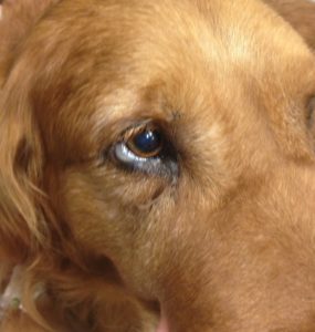
Effigy 3. Swelling of the conjunctiva without signs of inflammation or irritation is known as chemosis. This is a belatedly sign of fluid overload; it is incumbent on veterinary technicians to recognize earlier signs such every bit increased respiratory charge per unit and effort, increased breath sounds (eastward.g., crackles), or clear nasal discharge.
Fluid Types Bachelor
Several types of fluids are available, ranging from crystalloids to synthetic colloids to natural colloids (i.east., claret products). Each blazon has its place in the treatment of various conditions and pathologies constitute in veterinary patients. It is easiest to differentiate fluids based on their purpose: maintenance or replacement therapy. Table ane outlines the components of mutual maintenance and replacement fluids available to veterinary practitioners in the United States. The resources listed in the Recommended Reading box can provide more detailed explanations of fluid types and their effects.

Table 1. Composition of Common Veterinary Fluids
Crystalloids
Patients presented as an emergency often require firsthand intravascular expansion in the form of crystalloid boluses, or large volumes of crystalloid fluids. Crystalloid fluids move apace from the intravascular space into other fluid compartments, primarily the intracellular compartment. Less than 1-tertiary of the crystalloid volume administered intravenously persists in the vasculature 1 60 minutes subsequently administration,4 making these fluids an excellent choice for treating dehydration and electrolyte derangements and correcting gratuitous water deficits.
Crystalloid fluids can be categorized equally follows:
- Gratuitous water: 5% dextrose in sterile water or 0.45% saline. This hypotonic (i.e., containing fewer solutes than ICF) solution replenishes the interstitial fluid and ICF compartments.
- Replacement solutions: These balanced, isotonic solutions are designed to replenish the ECF compartments, including increasing intravascular volume and restoring perfusion. Isotonic fluids incorporate a solute concentration that approximates that of ICF, and crystalloids that are considered "replacement" fluids (TABLE one) have compositions that closely friction match the electrolyte residue and pH of ECF,1 making them platonic to supplant losses from that fluid compartment (e.one thousand., dehydration).
- Maintenance solutions: These balanced, isotonic solutions have less sodium and more potassium than replacement fluids and may be more suitable for long-term fluid therapy afterwards restoration of intravascular volume and correction of electrolyte derangements. Maintenance fluids are rarely used alone—they are commonly combined with a ratio of 0.9% sodium chloridei (aka "normal" or "isotonic" saline) to more closely lucifer the composition of the fluid in the intravascular space, preventing unwanted fluid shifts between compartments.
- Hypertonic solutions: 7% to 23.4% saline. These fluids comprise a solute concentration higher than that of ICF and quickly expand intravascular volume by drawing water from the interstitial and intracellular compartments. Because of this oncotic pull, hypertonic solutions should never be used in cases of astringent dehydration.
Colloids
Many practitioners likewise employ colloids (either synthetic or natural) in an emergency to expand the intravascular compartment without the risk of fluid overload posed by infusing large volumes of crystalloid fluids. Colloids contain big, osmotically agile particles that work to agree fluid in the vasculature after administration.
Constructed colloids are fluids with large molecules designed to provide oncotic pressure support within the intravascular space. Natural colloids are claret products such as whole blood, packed ruddy blood cells (pRBCs), plasma, and albumin. Whole blood and pRBCs have the added benefit of providing oxygen-carrying capacity, helping to forestall and care for hypoxia.
The utilize of colloids is highly controversial in human medicine and becoming so in veterinary medicine also,12 with recent research13 implicating a link between the use of a constructed colloid and the development of acute kidney injury in dogs.
Developing and Implementing a Fluid Therapy Plan
In that location is a helpful guideline when it comes to fluid therapy: Supplant like with like. This means if a patient has lost claret, that fluid should be replaced with plasma, pRBCs, or whole blood. If a patient has lost trunk fluids through diarrhea, vomiting, or excessive urination, replacement should be with similarly constituted isotonic crystalloid fluids. While development of the fluid plan is ultimately the veterinarian's purview, it is of import for veterinary nurses and technicians to sympathise the fluids available and for what conditions they might be used in clinical practise.
Fluid therapy in the veterinary infirmary or dispensary has three main phases, which tin overlap and alternate, depending on how a patient presents and the progression of its disease process. The resuscitation phase refers to correcting daze and other life-threatening fluid deficits; the replacement phase is the time taken to replace dehydration deficits; and the maintenance stage covers fluids provided during hospitalization to support and maintain homeostasis. BOX three provides examples of fluid choices in some specific disease processes.
BOX 3 Appropriate Fluid Choices for Selected Disease Processes
- Cardiac disease: Low-dose maintenance crystalloid, such every bit 0.45% saline with dextrose (may crave potassium and or magnesium supplementation)
- Airsickness/diarrhea: Replacement crystalloid, such as lactated Ringer's solution, Normosol-R, or
Plasmalyte-A - Diabetic ketoacidosis: Replacement crystalloid, such as lactated Ringer'southward solution, Normosol-R,
Plasmalyte-A - Hemorrhage: Natural colloid, such every bit plasma, whole blood, pRBCs
The amount of fluid to exist provided to a patient must exist calculated carefully, taking into business relationship the need for intravascular volume expansion, the profundity of perfusion deficits, the caste of dehydration, and the severity of electrolyte derangements, amongst other considerations. BOX 4 lists common fluid therapy calculation formulas.
BOX 4 Fluid Therapy Formulas
Calculation of Aridity Deficit i
Body weight (kg) × % dehydration equally a decimal = liters of fluid required to right dehydration
Calculation of Maintenance Fluid Requirements*
Dogs: Body weight (kg)0.75 × 132 = 24-hour fluid requirement in milliliters
Cats: Trunk weight (kg)0.75 × 80 = 24-hour fluid requirement in milliliters
Ongoing losses (e.g., from diarrhea, vomiting, or polyuria) must be calculated and added to the total maintenance requirement obtained from these formulas.
*UC Davis School of Veterinary Medicine fluid therapy formula.
Conclusion
Understanding the demand for fluid therapy, methods of providing fluids, types of fluids available, and how to keep patients safe while providing this vital treatment is a large part of beingness a veterinary technician. Become with the menses and help patients feel better!
References
- DiBartola SP, Bateman S. Fluid, Electrolyte, and Acid-Base Disorders in Small Animal Do. 3rd ed. St. Louis, MO: Saunders Elsevier; 2006.
- Creedon JM, Davis H. Catheterization of the venous compartment. In: Advanced Monitoring and Procedures for Modest Animal Emergency and Critical Care. Chichester, West Sussex, Uk: Wiley-Blackwell; 2012:51-68.
- Davis H, Jensen T, Johnson A, et al. 2013 AAHA/AAFP fluid therapy guidelines for dogs and cats. JAAHA 2013;49(three):149-159.
- Silverstein DC, Hopper Chiliad. Pocket-sized Animal Critical Intendance Medicine. 2d ed. St. Louis, MO: Saunders/Elsevier; 2015.
- Macintire DK, Haskins SC. Manual of Small Brute Emergency and Disquisitional Care Medicine. second ed. Philadelphia, PA: Lippincott Williams & Wilkins; 2005.
- Reddick Advertisement, Ronald J, Morrison WG. Intravenous fluid resuscitation: was Poiseuille right? Emerg Med J 2011;(28):201-202.
- Hackett TB, Mazzaferro EM. Professional intraosseous catheterization. In Veterinary Emergency and Critical Care Procedures. Ames, IA: Blackwell; 2006:263-267.
- Spivey WH, Malone D, Unger Hard disk drive, et al. Comparison of intraosseous, cardinal, and peripheral routes of administration of sodium bicarbonate during CPR in pigs. Ann Emerg Med 1985;14(v):514.
- Stack AM. Three. Intraosseous infusion. In: Wolfson AB, Wiley II JF, eds. Textbook of Pediatric Emergency Procedures. 2nd ed. Philadelphia, PA: Wolters Kluwer Health/Lippincott Williams & Wilkins; 2008:281-288.
- Wingfield WE, Raffe MR. Emergency vascular access and intravenous catheterization. In: The Veterinary ICU Volume. Jackson Hole, WY: Teton NewMedia; 2002:58-67.
- Mazzaferro EM. Fluid therapy: the critical balance between life and death. Clinician's Cursory 2006:73-75.
- Cazzolli D, Prittie J. The crystalloid-colloid debate: consequences of resuscitation fluid selection in veterinarian disquisitional care. J Vet Emerg Crit Intendance 20105;25(1):6-19.
- Hayes Thousand, Benedicenti L, Mathews K. Retrospective cohort study on the incidence of acute kidney injury and death following hydroxyethyl starch (HES x% 250/0.5/five:1) administration in dogs (2007-2010). J Vet Emerg Crit Care 2015;26(1):35-twoscore.
Recommended Reading
- Davis H, Jensen T, Johnson A, et al. 2013 AAHA/AAFP fluid therapy guidelines for dogs and cats. JAAHA 2013;49(iii):149-159.
- DiBartola SP, Bateman Southward. Fluid, Electrolyte, and Acid-Base of operations Disorders in Small Animal Practice. 3rd ed. St. Louis, MO: Saunders Elsevier; 2006. Chapters 14 and 23.
- Pre-Hospital Push. IV catheter size: How much of a difference does it make? http://www.prehospitalpush.com/2016/03/04/four-catheter-size-how-much-of-a-deviation-does-it-brand/. Accessed March 30, 2016.
Source: https://todaysveterinarynurse.com/internal-medicine/the-basics-of-fluid-therapy-for-small-animal-veterinary-technicians/
Posted by: duckwifeentent1998.blogspot.com
0 Response to "Which Laboratory Changes Would You Expect To See In A Dehydrated Animal"
Post a Comment